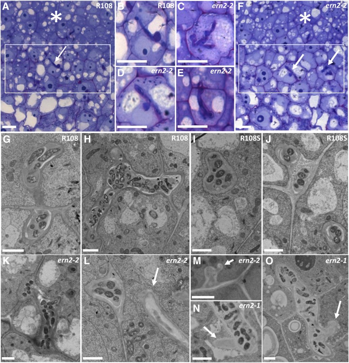
Figure 4.
The ern2 nodules are partially defective in IT development. Detailed views of infection zones (rectangles) of both wild-type R108 (A) and pink ern2-2 nodules (F) from toluidine-blue/basic fuchsin-stained 1-μm longitudinal sections. ITs within these regions (arrows) are highlighted in B (R108) and in C to E (ern2-2). ITs in ern2-2 are either similar to wild-type ITs or can exhibit a thickened appearance associated with less staining (C–E). Electron microscopy analysis has shown that some ITs of ern2-2 or ern2-1 can have zones devoid of bacteria (arrows in L–O) while others resemble wild-type ITs (compare R108 in G and H; R108S in I and J; ern2-2 in K–M; and ern2-1 in N and O). Nodule meristematic regions are highlighted by asterisks in A and F. Bars in A to F = 100 μm, G to O = 2 μm.

