Figure 8.
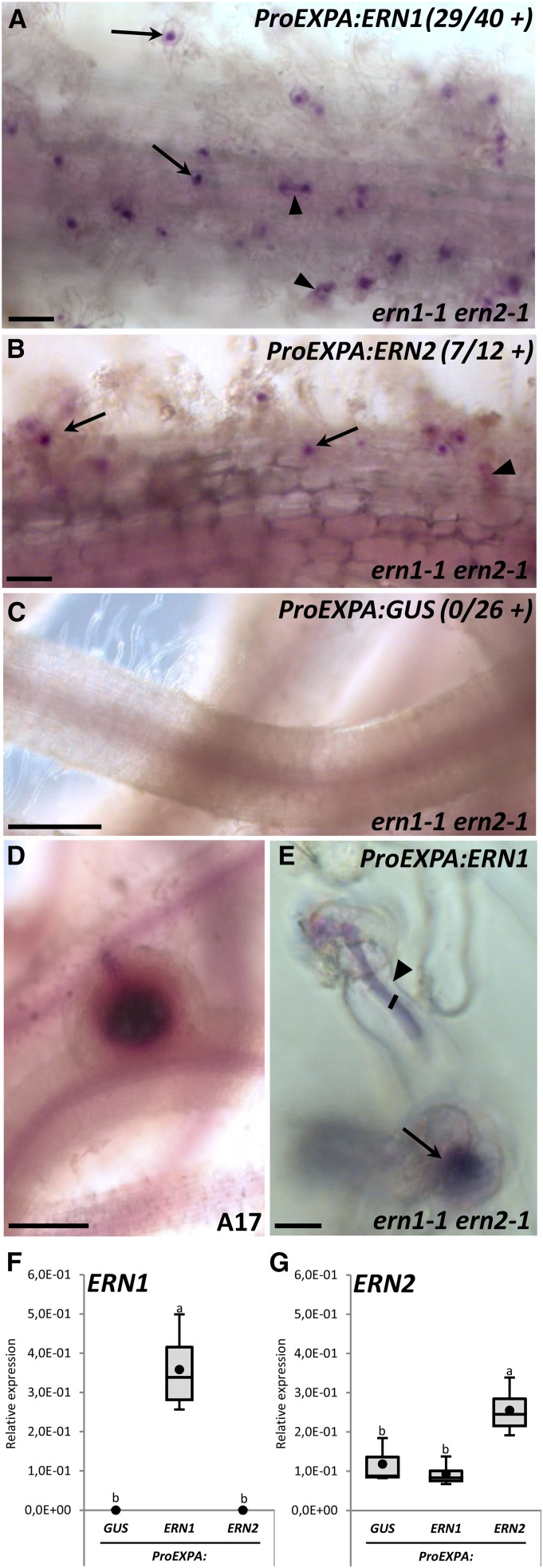
Epidermal expression of ERN1 and ERN2 can only restore RH infection. The ern1-1 ern2-1 double mutant was transformed by A. rhizogenes with a ProEXPA:ERN1 (A and E), ProEXPA:ERN2 (B), and ProEXPA:GUS negative control (C) constructs. Transformation of A17 with the ProEXPA:GUS construct was used as a positive control to validate nodule formation in the same experimental conditions (D). Transformed root systems were analyzed 3 to 4 wpi with S. meliloti-lacZ after staining for β-galactosidase bacterial activity (magenta staining). Typical “ern1-like” noninfected arrested nodules or infected nodules were not observed in any of the ern1-1 ern2-1 transgenic roots analyzed. Only RHCs or RH ITs (arrows and arrowheads in A, B, and E) were observed in complemented roots. Numbers in A to C represent the number of independent composite plants exhibiting restored RHC or RH infection from three (for ProEXPA:ERN1 and ProEXPA:GUS) and two (for ProEXPA:ERN2) independent experiments. F and G, QRT-PCR analyses in total root RNA samples from individual composite plants confirmed the expression of ERN1 or ERN2 in complemented roots. Values represent average of three to four independent plants after normalization against reference transcript levels. Black circles depict mean values. One-way ANOVA followed by a Tukey HSD test of the values were performed (in F, P < 0.001; in G, P < 0.01). Classes sharing the same letter are not significantly different. Bars = 100 μm (A and B); 500 μm (C and D); 10 μm (E).

