Figure 4.
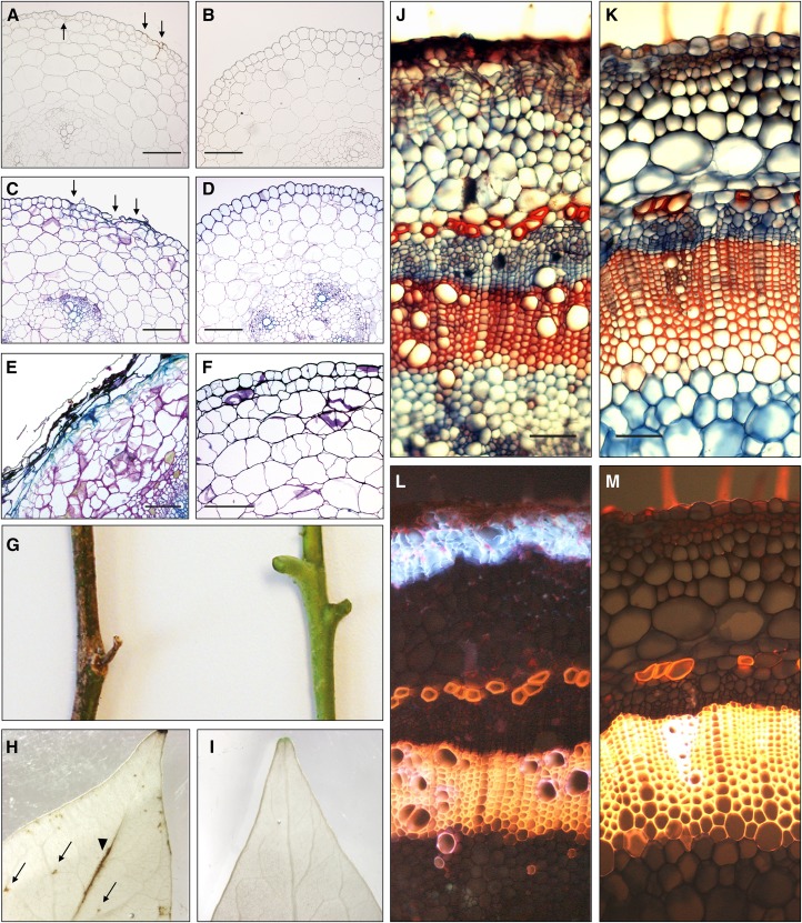
Sl2/3-MMP loss-of-function phenotypes. A to F, Hypocotyl cross sections (5 µm; 0.5 cm above the crown) of Sl2/3-MMP-silenced plants (A, C, and E) and wild-type plants (B, D, and F). The onset of cell death is indicated by arrows in unstained (A versus B) and Toluidine Blue-stained (C versus D) sections of 2-week-old seedlings. Deterioration of the epidermis, loss of cellular organization in the stem cortex, and accumulation of phenolic material as visualized by Toluidine Blue staining are apparent in 4-week-old seedlings (E versus F). Bars = 200 µm. G to I, Sl2/3-MMP loss-of-function phenotypes in fully grown tomato plants. G, Stem segments of 2-month-old HP and wild-type plants. H and I, Cell death in leaves. Destaining in boiling ethanol reveals necrotic lesions on top of the main vein (arrowhead) and within the leaf blade (arrows) in leaflets of silenced plants (H) but not in the wild type (I). J to M Formation of a secondary dermal tissue (wound periderm) in stems of fully developed tomato plants silenced for Sl2/3-MMP expression (J and L) compared with wild-type controls (K and M). The periderm is visible as distinct files of cells at the periphery of the stem section (hand cut, stained with safranin/Alcian Blue; J). In the same section, blue autofluorescence (excitation at 365 nm) indicates the accumulation of phenolic compounds in cell walls of suberized peridermal cork cells (L). Bars = 100 µm.

