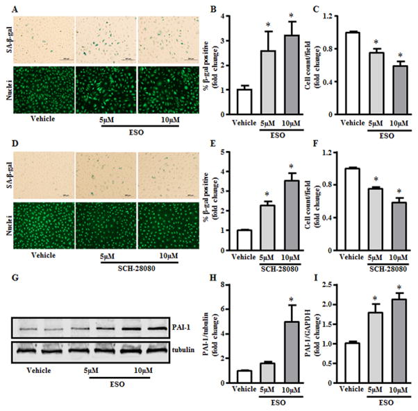
Figure 3. PPIs accelerate endothelial senescence.
(A&D) Senescent cell number detected by staining for senescence associated-β-galactosidase (SA-β-gal; upper panel) and for SYTO-13 to detect cell nuclei for total cell count (lower panel). (B,C,E and F) Respective quantification for % positive SA-β-gal cells and average cell count per field (n=6). (G and H) PAI-1 protein expression by Western blot analysis (n=3). (I) PAI-1 mRNA expression quantified by RT-PCR (n=6). *p< 0.05 vs vehicle (DMSO).

