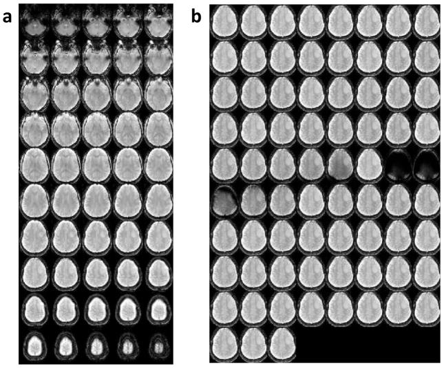
Figure 1.
A representative 3D whole-brain pulsed steady-state CEST image using low RF saturation power for a low-grade (grade II) oligodendroglioma patient. (a) 3D unsaturated images of the steady-state acquisition covering the whole brain. (b) A montage of a single slice image for saturation offset frequencies ranging from −18 to 18 ppm.

