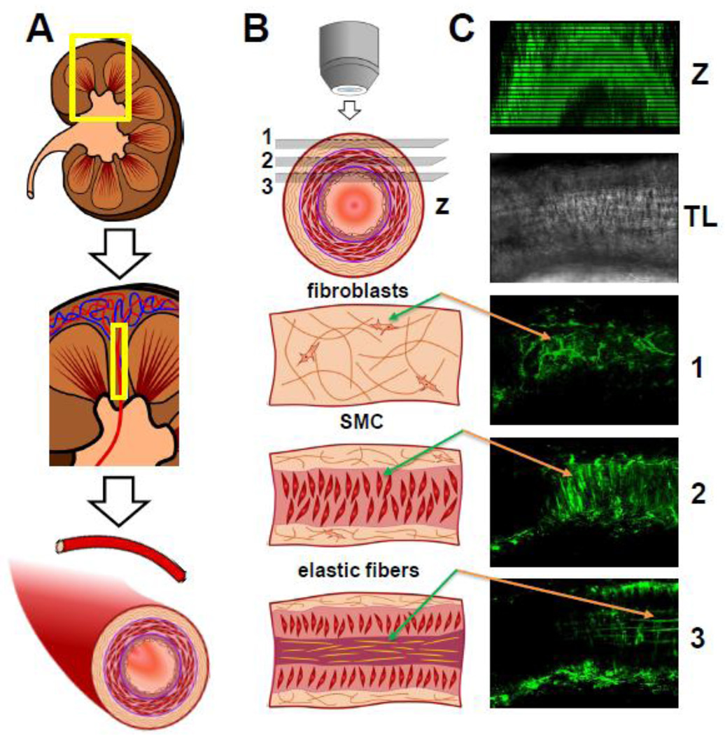
Fig. 2. The basic strategy for isolation and imaging of the renal vessels.
(A) Dissecting of renal resistance arteries. (B) Imaging renal vessels with two photon microscopy allows detecting three distinct tissue layers at different depths within the vessel wall: 1, fibroblasts rich in collagen; 2, smooth muscle cells (SMC) and 3, endothelial cells/elastic fibers. (C) Example of renal vessel imaging loaded with Fluo-4 AM Ca2+ indicator: Z-stack of the vessel, TL – vessel in transmitted light, 1, 2, and 3 corresponding layers are described in B.

