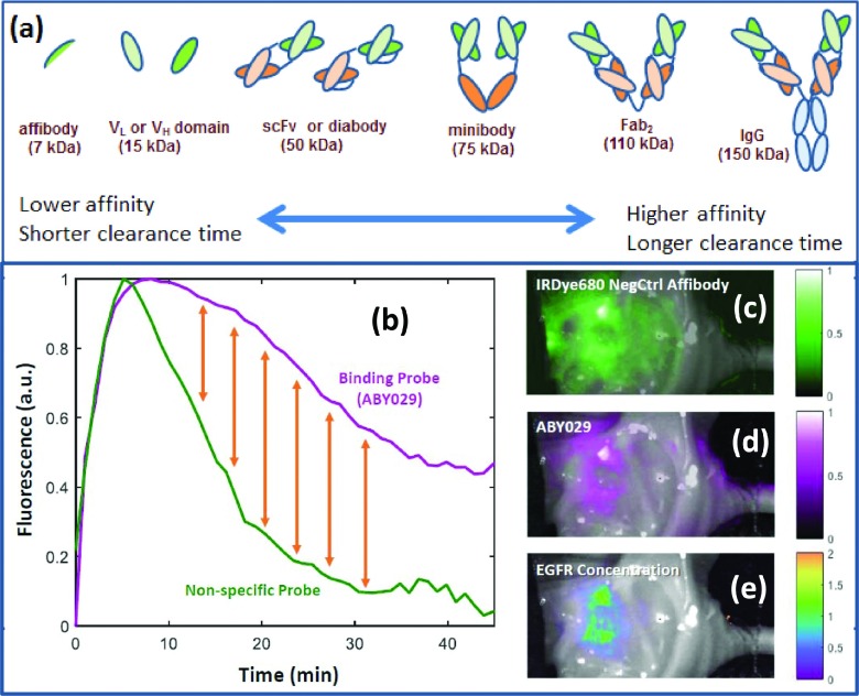
FIG. 5.
The range of immunological probes which could be utilized for cell surface receptors are shown (a), with the trade-off that comes with variation in size, being affinity for metabolic clearance time (Refs. 127 and 133). In (b) the problem of binding versus nonbinding probes is illustrated, in that even a specifically binding probe has significant signal simply from the wash in and wash out kinetics of perfusion. A nonspecific probe could be utilized to provide a reference, with the difference between the two being the bound fraction of specific probe (yellow arrows) (Ref. 132). In (c)–(e), an example tumor image is shown where EGFR-binding affibody labeled with IRDye 800CW (called ABY-029) is shown localizing (d) and a nonspecific affibody control (c). These were imaged simultaneously after a simultaneous injection, and the fitted difference signal between the two images shows the smaller EGFR + bound region (e).

