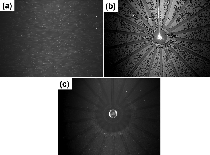
(a) Both blood cells and fluorescent nanocolloids were distributed randomly when no electric field was applied. (b) In a frequency range between 1 and 3 kHz, fluorescent nanocolloids were conveyed along the electrodes and concentrated onto the centre electrode, and blood cells were excluded away from the electrodes (Multimedia view). This image is enhanced as a video in the supplemental material. (c) When the applied frequency was below 1 kHz and above 3 kHz, the higher positive DEP and electrophoretic forces as compared to the ACEO drag caused the formation of concentrated nanocolloids in the form of a ring-shape. (Multimedia view) [URL:
http://dx.doi.org/10.1063/1.4953447.1]
Download video file (8MB, mpg) DOI: 10.1063/1.4953447.1