
Fig. 5.
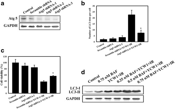
Measurement of autophagic and cytotoxic effects in 4 T1 cells pretreated with Atg shRNA. a Western blotting for Atg5. The cells were transfected with Atg5 shRNA for 24 h. b Quantitative data calculating the number of LC3 dots per cell in the absence or presence of Atg5 shRNA. c Cytotoxic effects in the absence or presence of Atg5 shRNA. Cells were transfected with Atg5 shRNA for 24 h and were then incubated with YCW1 (1 μM) and IR (4 Gy) for 48 h. *, p < 0.05, YCW1 + IR versus Atg5 shRNA + YCW1 + IR. Data are presented as the mean ± standard deviation of three independent experiments. d Western blot analysis of LC3-I and LC3-II expression in 4 T1 cells. Cells were pretreated with BAF for 1 h prior to IR treatment and then treated with YCW1 (1 μM) and IR (4 Gy) for 48 h