
Fig. 7.
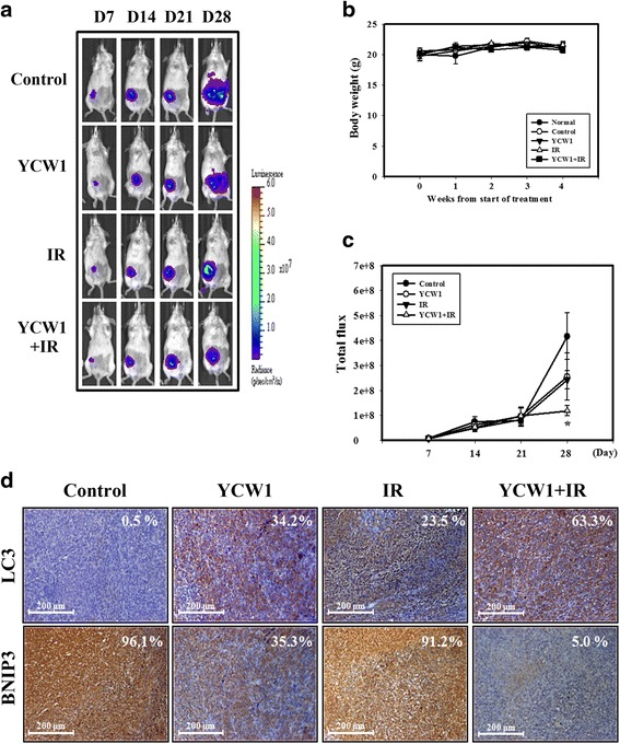
Combined treatment with YCW1 and IR enhances the anti-tumor effects in an orthotopic breast cancer model. a 4 T1-luc cells were injected into the mammary fat pads of Balb/c mice, observed for luciferase signals and photographed using an IVIS 200. b Measurement of body weight in Balb/c mice taken once per week. c Quantification of the luciferase signals. *, p < 0.05, versus control. d IHC staining of orthotopic tumor tissues from the mice. IHC was used to determine the expression levels of LC3 and BNIP3 (×100 objective magnification). The percentage of LC3 and BNIP3-positive cells was determined using HistoQuest software (TissueGnostics)