
Fig. 1.
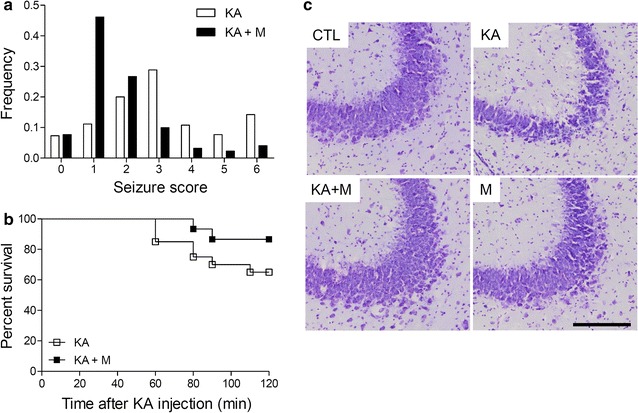
Mdivi-1 effects on KA-induced seizure activity, percent survival, and neuronal cell death. The frequency of behavioral seizure scores (a) and percent survival (b) were monitored during the first 2 h after KA treatment (P < 0.05 KA versus KA + M). c Cresyl violet-stained brain sections showing pyramidal neurons in the CA3 regions. The brain sections were prepared at 24 h after KA injection. Scale bar 200 µm