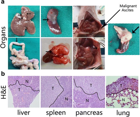
Fig. 4.

Macroscopic and microscopic examination of organs with metastatic lesions and orthotopic pancreatic tumours. 24 mice were randomly divided into three groups (PANC-EV, PANC-MUC4/Y, PANC-MUC4/Y-AMOPΔ). The different groups PANC-1 derived cells (2 × 10⁶ cells/100 μl) were orthotopic implanted into pancreas. Forties five dayed later, mice were euthanised by CO2 asphyxiation and autopsied (a) The macroscopic examination of organs with metastatic lesions (includes liver, spleen, lung, peritoneum, and mesentery), malignant ascites and orthotopic pancreatic tumours. The arrows pointed the metastatic nodes. b H&E staining was used to the histological change. Normal (N) and tumour lesions (T) in different organs are separated as indicated
