Fig. 2.

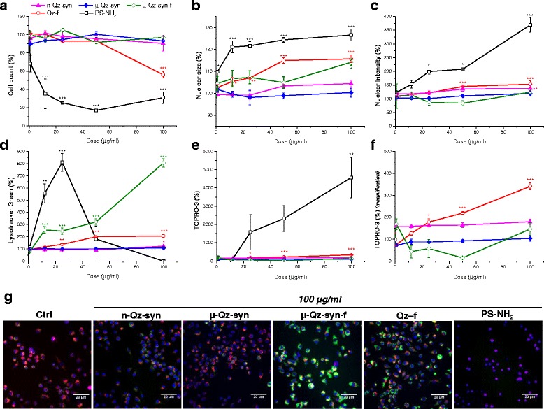
Cell toxicity in RAW 264.7 macrophages exposed to as-grown (n-Qz-syn and μ-Qz-syn) or fractured (μ-Qz-syn-f and Qz-f) quartz crystals. RAW 264.7 murine macrophages were exposed for 24 h to medium (Ctrl) or increasing concentrations of n-Qz-syn, μ-Qz-syn, μ-Qz-syn-f, Qz-f, and PS-NH2 beads (cytotoxic control). a Cell count (number of Hoechst stained nuclei), b nuclear size (average object area of Hoechst), c nuclear intensity (Hoechst intensity), d lysosomal acidification (Lysotracker Green intensity), and e plasma membrane integrity (TOPRO-3 intensity) were measured. A magnification from 0 to 500 % of TOPRO-3 intensity plot is given in panel f. As-grown quartz crystals (n-Qz-syn and μ-Qz-syn) were inactive for all the cytotoxicity parameters investigated, while fractured quartz (μ-Qz-syn-f and Qz-f) induced significant cell stress. Data are reported as mean percentages of the control ± SEM in a representative experiment performed in triplicate. *p < 0.05, **p < 0.01 and ***p < 0.001 vs control not exposed to quartz. Representative images captured by epifluorescence microscopy on RAW 264.7 macrophages exposed to Ctrl or quartz samples at 100 μg/ml (g). Blue fluorescence is indicative of nuclear staining, green fluorescence of acidic compartment staining, red fluorescence of mitochondrial membrane potential, and violet fluorescence of cell membrane permeability. Scale bars = 20 μm
