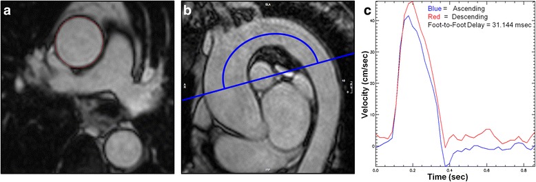
Fig. 1.

a Ascending aortic cross-sectional measurements made by manual planimetry of the aortic endovascular-blood pool interface at minimal and maximal distension. b Sagittal oblique CMR image from which the length of the aortic arch is manually measured. The image is subsequently used to determine site of acquisition of phase contrast cines. c Time-Velocity curve derived using PMI software to calculate foot-foot delay (curves are automatically adjusted/overlaid to accommodate time delay)
