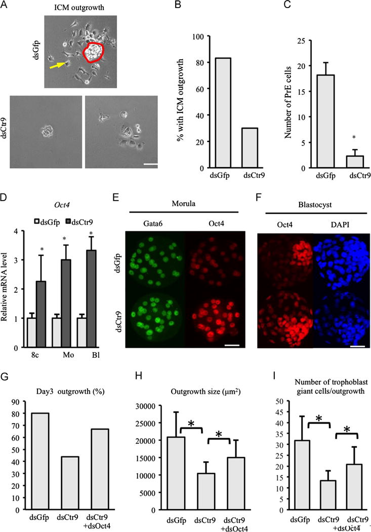
Fig. 3.

Depletion of CTR9 abrogates ICM identity and differentiation potential. 3 days in culture following immunosurgical removal of TE, primitive endoderm cells (yellow arrow upper panel in A) migrate away from control ICM (red circle, upper panel in A). Very few cells (either ICM or PE) are present when Ctr9-deficient ICMs are plated in culture (A, lower panels). Scale bar: 20 μm. (B) Graph representing outgrowth potential of isolated ICM. (C) Quantification of the number of primitive endoderm cells observed per ICM. (D) Oct4 mRNA abundance is increased consistently beginning at the 8-cell after Ctr9 knockdown. (E,F) OCT4 protein level is enhanced in morula (15 dsGfp, 14 dsCtr9 morula examined) and trophectoderm cells of blastocysts (16 dsGfp, 25 dsCtr9 blastocysts examined) in dsCtr9 embryos. Scale bar: 50 μm. (G–I) Reduction of Oct4 partially rescues dsCtr9 outgrowth potential (G), average outgrowth size (H) and number of trophoblast giant cells observed (I, 5 dsGfp, 9 dsCtr9, 6 dsGfpþdsOct4 embryos examined). Asterisks indicate P<0.05 by student T-test. Error bars represent SEM.
