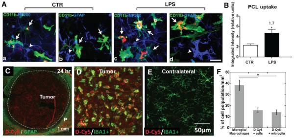
Figure 4. Specific uptake of nanoparticles by microglial cells and tumor-associated microglia/macrophage.
(A) Representative confocal image of PCL nanoparticles uptake by control (non-activated) microglia LPS stimulated (activated) microglia in vitro, (B) Semi-quantification based on integrated fluorescence intensity demonstrated higher 1.7-fold higher uptake of particles by LPS stimulated microglia than control microglia. Reprinted (adapted) with permission from Ref [92]. Copyright (2015) American Chemical Society. (C–F) Distribution of fluorescent-labeled generation 4 hydroxyl terminated PAMAM dendrimers (D-Cy5) in a rat brain bearing 9L glioblastoma. D-Cy5 was administrated systemically at day 9 after tumor inoculation. Confocal images of tumor region suggested that D-Cy5 (~4 nm in size) uniformly distributed across the 6 mm tumor after 24 hr post administration. (C), and was co-localized with Iba1+ TAM in tumor but not in the 'resting' microglia in the contralateral hemisphere of the same rat brain after 4 hr of D-Cy5 administration (D and E). Imaging analysis showed almost all D-Cy5+ cells were Iba1+ cells, indicating the high efficiency of TAMs taking up dendrimers (F) [101]. Green: Iba1+ microglia/macrophages; red: D-Cy5; blue: DAPI; bar: 100 μm. Reprinted from Biomaterials, Vol 52, F Zhang, et al., Uniform brain tumor distribution and tumor associated macrophage targeting of systemically administered dendrimers, 507-516, Copyright (2015), with permission from Elsevier.

