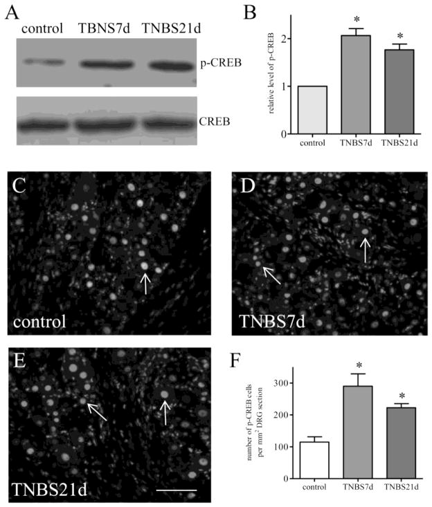
Figure 1. CREB activity was increased in the DRG post colitis.
Western blot of phospho-CREB (A) was performed to identify CREB activity level in the whole DRG (pooled L1–L2 and L6–S1) showing increases (B) at both 7 days and 21 days post colitis induction (animal numbers: control, 6; TNBS 7 days, 6; TNBS 21 days, 5). Immunostaining of phospho-CREB (C–E) demonstrated that phospho-CREB was expressed in the nucleus of DRG neurons (arrows). Number counting of phospho-CREB in DRG neurons (F) showed significant increases at 7 days and 21 days post colitis induction (animal numbers: control, 4; TNBS 7 days, 4; TNBS 21 days, 3), confirming the western blot results above. *, p<0.05. Calibration bar = 40 μm. Representative microphotographs (C–E) are from L1 DRGs.

