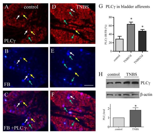
Figure 3. PLCγ expression was increased in bladder afferent neurons post colitis.
Immunostaining showed that PLCγ expression (A and D, red cells) was expressed in bladder afferent neurons (B and E, blue cells) in both control animals (A–C) and animals treated with TNBS (D–F). PLCγ expression was increased in bladder afferent neurons (compare F to C, purple cells indicated by green arrows) at 7 days and 21 days post colitis induction (G, animal numbers: control: 5, TNBS 7 days, 4; TNBS 21 days, 4). Western blot confirmed that PLCγ expression was up-regulated in the DRG by colitis (H, n=3). *, p<0.05. Calibration bar = 80 μm.

