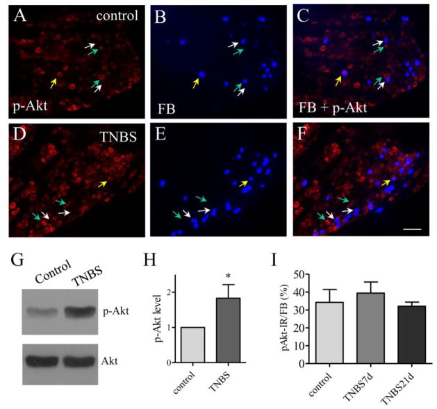
Figure 4. The expression level of phospho-Akt was not changed in bladder afferent neurons post colitis.
Immunostaining showed that a small population of phospho-Akt immunoreactivity (A and D, red cells) was expressed in bladder afferent neurons (B and E, blue cells) in both control animals and animals treated with TNBS (compare F to C). Western blot showed an up-regulation of Akt phosphorylation (activation) in the DRG post colitis (G and H, n=3). However, the number of bladder afferent neuron expressing phospho-Akt expression was not changed at 7 days and 21 days post colitis induction (I, animal numbers: control: 5, TNBS 7 days, 4; TNBS 21 days, 4). Calibration bar = 60 μm. *, p<0.05.

