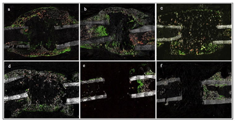
Fig. 10.
Representative fluorescent images at 14 days postoperatively. Mineralized woven bone spanning the length of the defect is evident in the rhBMP-2 (a), rhBMP-2 + OPG (b), LV-BMP-2 (c) and LV-BMP-2 + OPG groups (d), with the bony bridge being more robust in the BMP-2 + OPG treated femora (b, d). A prominent cellular response, with numerous mature osteoblasts present in the defect site, was observed for all BMP-2 treated groups (a–d). In the two BMP-2 alone groups, the bony bridge consisted mostly of GFP+ cells with more modest amounts of bone in comparison with the corresponding BMP-2 + OPG groups. The carrier alone (e) and OPG alone (f) controls exhibited minimal bone formation, with GFP+ cells limited to the endosteum, intramedullary canal and within the newly formed bone on top of the original cortex (periosteal reaction).

