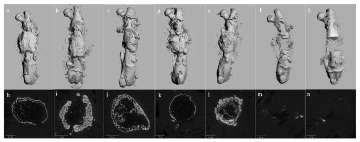
Fig. 3.
Representative micro-CT images obtained in the longitudinal and axial planes showing the healing of the femoral defect at the 56-day time point. Complete healing of the defect is evident in the longitudinal view for the BMP-2 treated groups (a. rhBMP-2, b. rhBMP-2 + OPG, c. rhBMP-2 + delOPG, d. LV-BMP-2, e. LV-BMP-2 + OPG). The axial view (h–l) indicates the formation of new cortices and reconstitution of the medullary canal across the femoral defect. A thicker cortex is observed in BMP-2 + OPG treated groups (i, j, l) compared to the groups treated with BMP alone (h and k). Minimal bone formation within the femoral defect is seen in the carrier alone (f and m) and OPG alone (g and n) groups.

