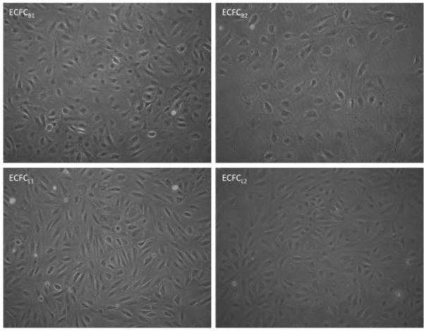
Figure 1. Phase images of ECFCB and ECFCL clones.

Confluent monolayers of each ECFC clone expanded from single cell isolates from human whole blood are shown. Magnification is 40x.

Confluent monolayers of each ECFC clone expanded from single cell isolates from human whole blood are shown. Magnification is 40x.