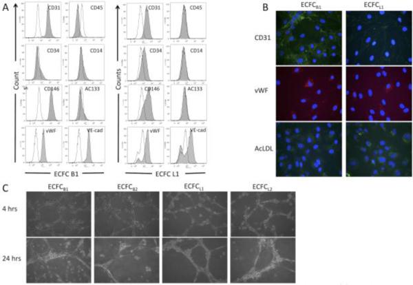
Figure 2. Isolated clones express endothelial markers, uptake Ac-LDL, and undergo capillary morphogenesis.
(A) ECFC clones B1 and L1 were stained for endothelial or monocyte/macrophage markers and the expression was analyzed by flow cytometry. Shaded areas are primary antibody, unshaded areas are IGG control. (B) ECFC clones B1 and L1 were stained for CD31 (green) or von Willebrand Factor (red) or incubated with fluorescent-labeled AcLDL (green). Nuclei in all samples were stained with DAPI (blue). (C) ECFC clones B1, B2, L1, and L2 were plated on Matrigel. Cells were monitored for capillary morphogenesis and photographs were taken at the indicated times. These experiments were each performed 3 separate times with identical results.

