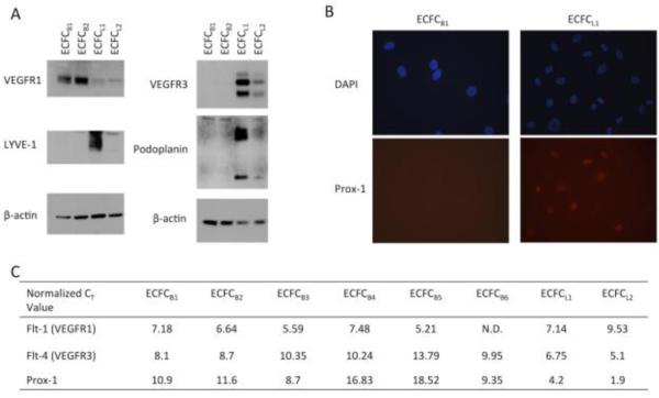
Figure 3. Expression of lymphatic-specific markers in ECFC clones.
(A) ECFC lysates were separated on a 4-20% SDS-PAGE gel and transferred to PVDF membrane. Blots were probed for VEGFR-1, VEGFR-3, LYVE-1, podoplanin, or β-actin. (B) ECFC B1 and ECFC L1 were stained for Prox-1 protein expression (red) and DAPI (blue). Each experiment was performed 3 times with similar results. (C) RNA was isolated from each ECFC clone. Real-time RT-PCR was performed for VEGFR-1, VEGFR-3, and Prox-1. Values shown are CT values of each gene normalized to the level of GAPDH in each sample.

