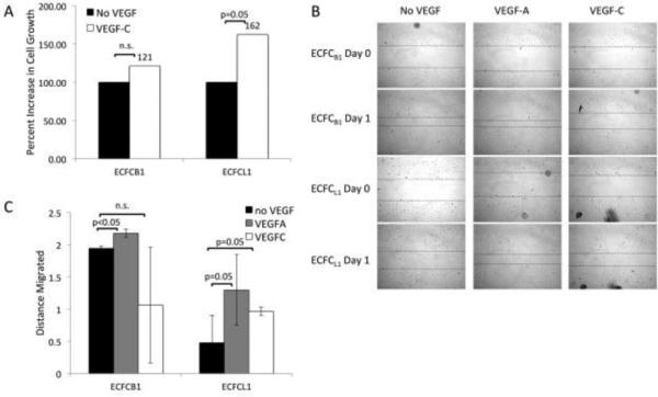
Figure 4. ECFC response to VEGF-A and VEGF-C.
(A) Cells were plated without VEGF (black bars) or with VEGF-C (white bars) for 4 days and then cell number was quantified with a hemocytometer. (B) Confluent monolayers of ECFC B1 or ECFC L1 were wounded and treated with or without VEGF-A or VEGF-C for 24 hours. Pictures were taken at the time of wounding and 24 hours later. (C) The distance migrated into each wound was quantified and graphed. Data shown is representative of 5 experiments.

