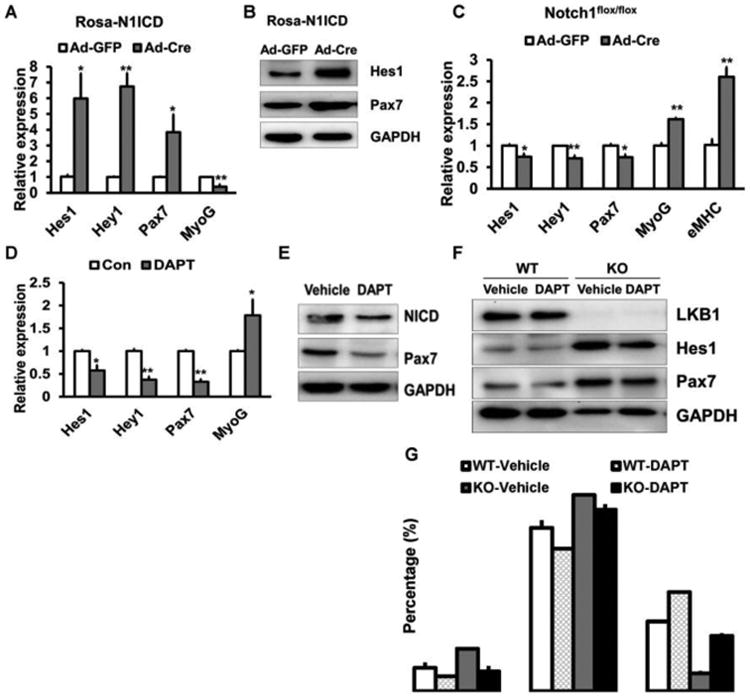
Figure 3. Lkb1 regulates Pax7 expression and myoblast division through Notch signaling pathway.

(A, B) The mRNA (A, n=5) and the protein (B) levels of Pax7 in Rosa26-N1ICD myoblasts infected with adenovirus-GFP or adenovirus-Cre.(C) Expression of Pax7 in Notch1flox/flox myoblasts infected with adenovirus-GFP or adenovirus-Cre (n=6). (D, E) Relative mRNA (D, n=6) and protein (E) levels of Pax7 in WT myoblasts treated with Notch inhibitor DAPT. (F) Protein levels of Pax7 in WT and MyoD-Lkb1 myoblasts treated with or without DAPT. (G) Percentages of self-renewal, proliferative and differentiation divisions in the WT and Lkb1 KO myoblasts treated with or without DAPT. n=3 independent experiments, with at least 50 doublets analyzed in each experiment. Error bars represent SEM, *P<0.05, **P<0.01
