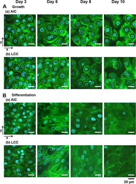
Fig. 3.
Different development of tight junctions in cell-to-cell contacts in various culture conditions. Actin filaments in NHBE cells grown in different culture conditions were stained with Alexa Fluor® 488 phalloidin (green) and cell nuclei stained with HOE (blue) in HBSS. (A) NHBE in the growth medium under (a) AIC or (b) LCC condition revealed imperfect tight junctions. (B) NHBE in the differentiation medium has developed intact tight junctions under either of (a) AIC or (b) LCC condition. Two-dimensional confocal images were displayed with the arrows for x- and y-axes. The scale bar below indicates 20 μm.

