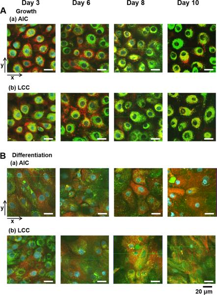
Fig. 4.
Two-dimensional (2D) architectures of NHBE cells on porous supports by confocal fluorescent imaging. Suborganelles (Cell nuclei, mitochondria or lysosomes) in NHBE cells grown in different culture conditions were stained with HOE (blue), MTR (red), and LTG (green) after 30 min-transport experiments with dye mixtures. (A) NHBE in the growth medium under (a) AIC or (b) LCC condition. (B) NHBE in the differentiation medium under (a) AIC or (b) LCC. Two-dimensional confocal images were displayed with the arrow indications (x- or y-axis). The scale bar below indicates 20 μm.

