
Fig. 5.
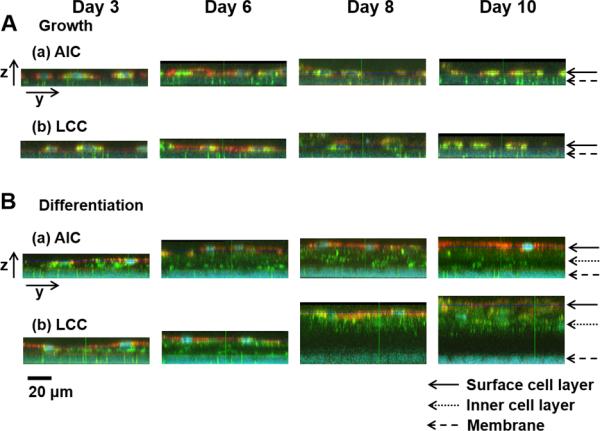
Various layer thicknesses of NHBE cells on porous membranes in inserts during the cultures (NHBEs in the (A) growth and (B) differentiation medium in (a) AIC or (b) LCC condition). In the images of yz planes corresponding to the 2D images of xy planes in Fig. 4, (B) NHBE cells in the differentiation medium formed multilayers with varied thickness of layers while (A) NHBEs in the growth medium showed monolayers with less intact between the cells, decreasing cell densities as a longer period of cultures. The scale bar indicates 20 μm. Cells in the surface or inner layers and porous membranes were indicated by the arrows.