Fig. 6.
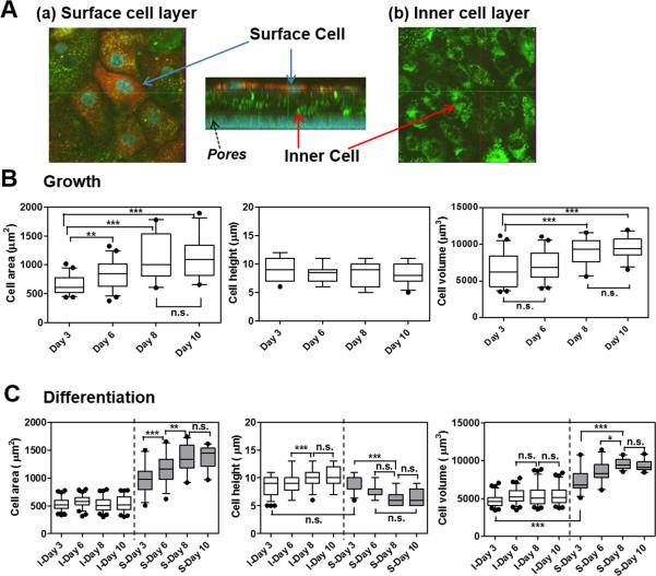
Variations in morphological parameters of NHBE cells on different days of AIC cultures. (A) Representative 3D view of the confocal image (z-stack) of NHBE in the differentiation medium on day 8. (a) Surface, (b) inner cell layers and pores of the membrane are indicated with the arrows. From the confocal images, morphometric parameters such as cell area, height, and volume were measured by MetaMorph software for the NHBE cells in the (B) growth medium; (C) differentiation medium on day 3, 6, 8, and 10 under air-interfacing condition. (C) In the case of NHBE cell multilayers in differentiation medium, inner (I) and surface (S) cell layers (separated with the dotted line) showed differences in cell area, height, and volume. Box-and-whisker plots showing median and 5-95 percentiles were depicted with statistical analyses. Statistics was performed with Tukey's multiple comparison test with the 5% significance level. n.s.=not significant, *P<0.05, **P<0.001, ***P<0.0001.

