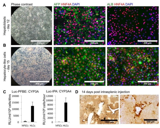
Figure 4. Hepatic specification and maturation of DE cells passed and differentiated in wells of a 96-well plate.
(A) After 8 days of hepatic specification (day 12), the cells were confluent, but did not display a hepatocyte morphology (phase contrast, magnification: 10x). Cells were positive for HNF4A and AFP, but remained negative for albumin, as assessed by immunofluorescence (Magnification: 20x).
(B) After hepatic maturation (day 15), hepatic-like cells (HLCs) displayed typical polygonal morphology (phase contrast, magnification: 10x) and were positive for AFP, albumin and HNF4A (magnification: 20x).
(C) Activity of the cytochrome P450 3A family (Substrate: Luc-PFBE) and CYP3A4 isoform (Substrate: Luc-IPA) in HLCs compared to hPSCs.
(D) Hep-Par1 immunohistochemistry visualization of engrafted HLCs 14 days after intrasplenic injection of 4 million HLCs in the spleen of MUP-uPA/SCID/Bg mice.
See Supplemental Figure 3 for more functional characterization of HLCs and assessment of intra-hepatic engraftment.

