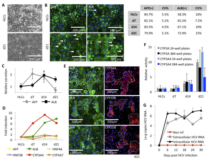
Figure 6. Long-term maintenance of HLCs.
(A) Improved phenotypic stability when HLCs are cultured in the new WEM hepatic maintenance medium, compared to previously described L15 medium.
(B) Immunofluorescence assays for expression of AFP and ALB in HLCs, differentiated in 384-well plates, and maintained for 3 weeks in the WEM medium. (Arrows indicate binucleated cells).Table indicates percentage of positive cells at the different times of culture in WEM medium, assessed by automatic quantification, and their respective coefficient of variation (CV%) from well to well.
(C) Relative secretion of AFP and ALB assessed by ELISA in the supernatants of culture of HLCs, differentiated in 384w plates, and maintained for 3 weeks in the WEM medium.
(D) RTqPCR analysis of hepatic genes in SC-derived HLCs, differentiated in 24-well plates, and maintained for 3 weeks in the WEM medium. See Supplemental Figure 2 for expression relative to control PHHs.
(E) Co-immunostaining for ALB, AFP, CYP3A4 and CYP2D6 in HLCs, differentiated in 24-well plates, after 21 days in WEM medium.
(F) Increased of the CYP3A PFBE and CYP3A4 IPA activities during 3 weeks of culture in WEM medium, on HLCs differentiated and maintained in 24-well plates (grey and light blue bars) and 384-well plates (black and blue bars).
(G) HCV replication in HLCs maintained in WEM medium for up to 1 month, assessed by RTqPCR for intra- and extracellular HCV RNA quantification, expressed respectively as log (copies HCV RNA / mg total cellular RNA) (black line) and log (copies HCV RNA / ml of supernatants) (grey line), compared to non-inoculated HLCs (red line). HLCs were differentiated, inoculated with HCVcc and maintained in 24-well plates.

