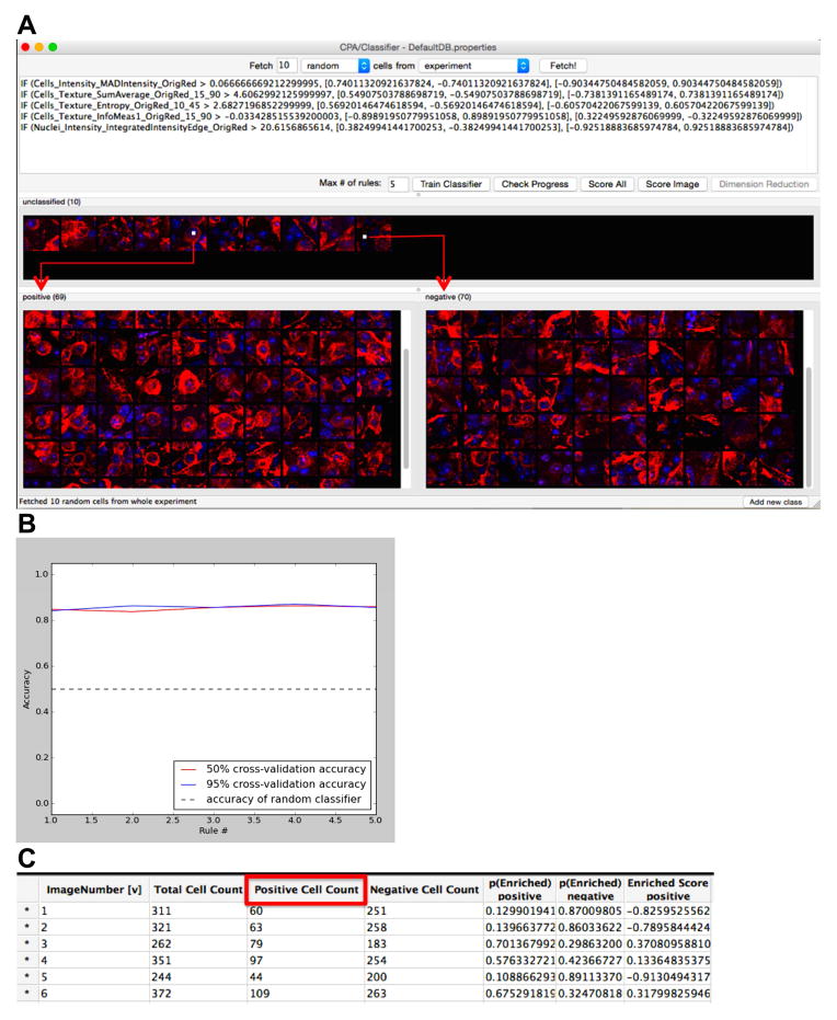
Figure 1. Automated scoring of cell morphologies using the CellProfiler Analyst machine learning and iterative feedback system.
(A) The software system presents the researcher with individual cells to classify from segments of images, sampled randomly from the set of images. After classification of a few cells, the researcher begins the iterative machine-learning phase (“Train Classifier”), in which the software generates a tentative rule based on the classified cells and presents the researcher with cells classified according to that rule. (B) The accuracy of the CPA classifier rule may be checked after each training set (“Check Progress”) and accuracy above 80% is considered suitable. (C) Following 4 training iterations, all cells from the experiment of interest are classified in order to calculate the number of positive (RGC) and negative (non-RGC) cells. “Positive Cell Count” denotes the cells recognized as RGCs. “Negative Cell Count” refers to image regions, in which the cells’ morphological features were not recognized as RGCs. “Total Cell Count” refers to the total number of cells recognized per image. “Image Number” represents the number of the image associated with the cell counts.

