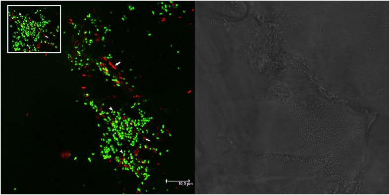
FIGURE 2.
Nodules may be colonized by different strains. Confocal laser scanning microscopy (CLSM) image of a portion of root nodule containing both BM687 (S. meliloti 1021 pBHR mRFP, in red) and BM286 (S. meliloti BL225C pHC60, in green) strains. Data from plate experiment. The bright field layer and the CLSM layer are shown. Arrows in the CLSM panel indicate both strains (in red and in green) colonizing the nodule. The inset highlights the central part of the CLSM image with BM687 and BM286 strains.

