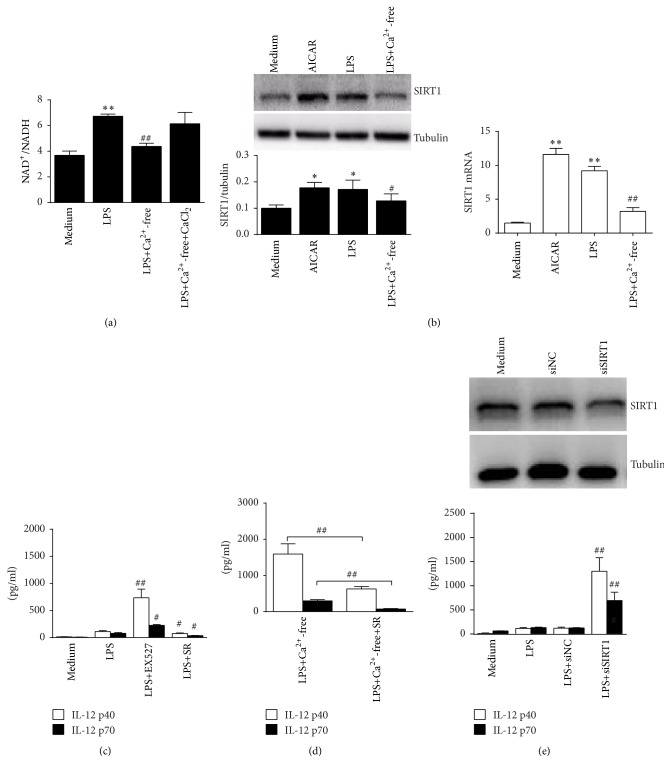
Figure 6.
SIRT1 is upregulated by AMPK activation to suppress IL-12 expression in the LPS-activated RAW 264.7 cells. (a) Cells were treated with 100 ng/mL LPS in normal DMEM or in DMEM without calcium or supplemented with 2 mM CaCl2 for 1 h. NAD+/NADH levels were detected (n = 3). (b) Cells were treated with 1 mM AICAR, 100 ng/mL LPS, or LPS plus calcium-free DMEM for 2 h. Protein and mRNA levels of SIRT1 were detected by western blot or real-time PCR. ∗ P < 0.05 and ∗∗ P < 0.01 versus medium; # P < 0.05 or ## P < 0.01 versus LPS (n = 3). (c, d) Cells were treated with LPS alone or with 2 μM EXT527 or 2 μM SRT1720 (c). Cells were also treated with LPS, calcium-free LPS, or calcium-free LPS with SRT1720 (d). Supernatants IL-12 p40 and IL-12 p70 (24 h) were detected by ELISA. # P < 0.05 and ## P < 0.01 versus LPS for each cytokine (n = 3). (e) Cells were treated with NC siRNA or SIRT1 siRNA for 24 h. Protein levels of SIRT1 were detected by western blot (upper). The siRNA-treated cells were further stimulated by 100 ng/mL LPS for 24 h (lower). Supernatants IL-12 p40 and IL-12 p70 were detected. # P < 0.05 and ## P < 0.01 versus LPS for each cytokine (n = 3).

