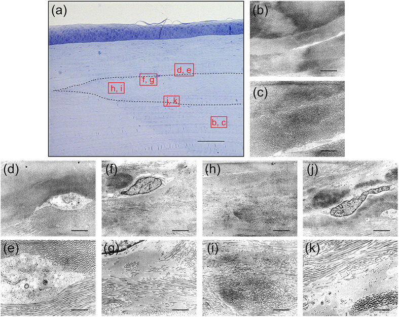
Figure 7. Structural analysis of decellularized corneas at 6 months after microkeratome-assisted anterior lamellar keratoplasty.
Optical micrograph of the decellularized cornea stained with toluidine blue at 8 weeks after interlamellar keratoplasty (a). The TEM images of the indicated squares in A are shown in (b–k). The dotted line region indicates the transplanted decellularized cornea. TEM observation indicated that the collagen fibrils structure of the decellularized cornea was highly organized and similar to the host corneas (d,e,h,i). An irregular collagen arrangement and an increase of spacing between the collagen fibrils remained (f,g,j,k). Scale bars: 100 μm in (a); 1 μm in (b,d,f,h,j); and, 3 μm in (c,e,g,i,k).

