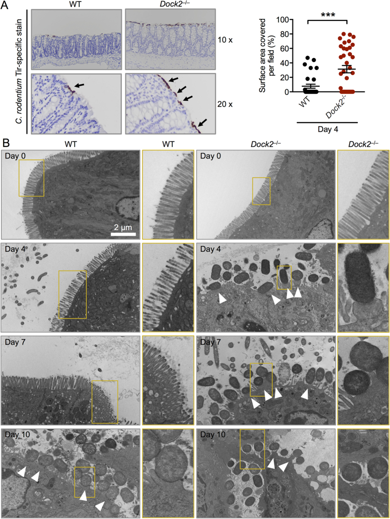
Figure 5. DOCK2 impairs bacterial attachment and formation of A/E lesions by C. rodentium.
(A) WT and Dock2−/− mice were orally infected with 1 × 10¹⁰ CFU of C. rodentium. Intestinal tissue sections were stained with anti-C. rodentium Tir antibody. Percentages of the area of the distal colon surface positive for Tir staining per field in infected WT mice (n = 5) and infected Dock2−/− mice (n = 6). Six different fields for each mouse were quantified. (B) Transmission electron microscopy images of the colonic intestinal epithelium of WT and Dock2−/− mice on Day 0 (uninfected), Day 4, Day 7 and Day 10 post-infection with C. rodentium. Arrowheads indicate bacterial attachment and formation of A/E lesions. Data are representative of two independent experiments (mean and SEM). Two-tailed t-test. ***P < 0.001.

