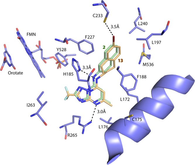
Figure 2.

X-ray structure of PfDHODH bound to 13 (PfDHODH-13). Limited residues from the 4 Å shell around 13 are shown, and the structure has been aligned to the PfDHODH structure bound to 2 (PDB 3I65) to allow comparison of the binding modes. Only the inhibitor 2 from 3I65 is displayed. PfDHODH amino acid, FMN, and orotate carbons are shown in purple, the carbons of 13 are shown in tan, and the carbons of 2 are shown in green. Nitrogens are blue, oxygens are red, sulfur is light yellow, fluorines are light blue, and bromine is deep red. Protein residues are labeled with their amino acid number.
