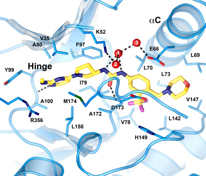
Figure 5.

Crystal structure of compound 20 (gold carbons) at the CDK8 active site. The DMG loop (center, D173 and M174 shown) is in the “out” conformation. A network of three solvent molecules (red, numbered spheres link the center of the compound to the protein). Water 2 makes an additional H-bond to the backbone carbonyl of D173 that was omitted for clarity. The hinge and C-helix (αC) are labeled.
