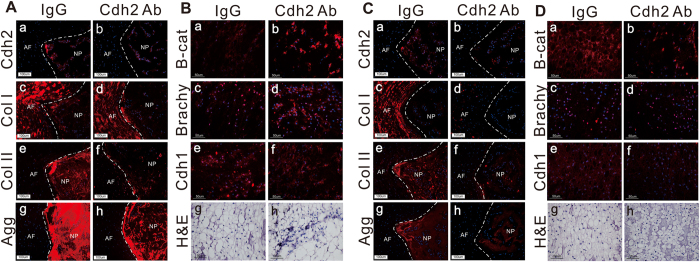
Figure 5. Delivering CDH2 blocking antibody in vivo results in loss of juvenile NP cell phenotype.
(A) Representative immunostaining of CDH2 (a,b), type I collagen (Col I) (c,d), type II collagen (Col II) (e,f), and aggrecan (agg) (g,h) in rat tail discs 2 weeks after intradiscal injection of CDH2 blocking antibody (Cdh2 Ab) or control immunoglobulin (IgG) (red = protein, blue = cell nuclei, scale bar = 100 μm, NP = nucleus pulposus; AF = anulus fibrosus). (B) Representative immunostaining of non-phosphorylated β-catenin (a,b), brachyury (c,d), and E-cadherin (e,f) and representative histological assessment of H&E staining (g,h) 2 weeks after intradiscal injection of Cdh2 Ab or IgG (red = protein, blue = cell nuclei, scale bar = 50 μm, NP tissue only). (C,D) Similar to panel A and B, but assessed 8 weeks after blocking antibody delivery.

