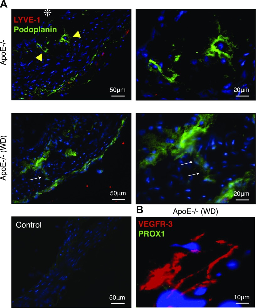
Figure 2.
Podoplanin+ LYVE-1− tube-like structures in atherosclerotic lesions. Double immunofluorescence staining for lymphatic endothelial markers, LYVE-1 (red) and podoplanin (green) in atherosclerotic lesions of apoE−/− mice. A) In aged apoE−/− mice fed normal diet (n = 5; 54–58 wk; M:F, 0.6) and in WD-fed apoE−/− mice (n = 3; 32 wk; M:F, 0.4), podoplanin+ LYVE-1− tube-like structures were found in the intimal space (yellow arrowheads). Podoplanin+ LYVE-1− tube-like structure crossing the intimal media border (white arrows). B) Immunofluorescence staining for the lymphatic endothelial markers, VEGFR-3 (red) and PROX1 (green). Intimal lymphatic-like structures expressed VEGFR-3 but not PROX1.

