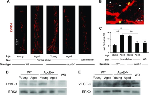
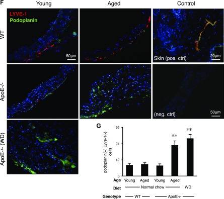
Figure 4.
Adventitial lymphangiogenesis. A) Whole-mount immunofluorescence staining for LYVE-1+ lymphatic vascular structures (red). Adventitial vessels are visualized in abdominal aortas (young WT mice: n = 5; 8–12 wk; M:F, 0.6; aged WT mice: n = 5; 54–58 wk; M:F, 0.6; young apoE−/− mice: n = 5; 8–12 wk; M:F, 0.6; aged apoE−/− mice: n = 5; 54–58 wk; M:F, 0.6; WD: n = 4; 32 wk; M:F, 0.5). Micrographs represent mosaics that were created by merging adjacent photomicrographs by using ImageJ. B) Photomicrograph of a representative atherosclerotic aorta illustrating LYVE-1+ lymphatic capillaries surrounded by LYVE-1+ macrophages (32-wk-old male, WD-fed apoE−/− mouse). C) Quantification of the LYVE-1+ areas as a percentage of the total aortic flatmount areas (young WT mice: n = 5; 8–12 wk; M:F, 0.6; aged WT mice: n = 5; 54–58 wk; M:F, 0.6; young apoE−/− mice: n = 5; 8–12 wk; M:F, 0.6; aged apoE−/− mice: n = 5; 54–58 wk; M:F, 0.6; WD: n = 4; 32 wk; M:F, 0.5). D) Western blot analysis of LYVE-1 in aortic walls, ERK-2 loading control (young WT mice: n = 4; 8–12 wk; M:F, 0.5; aged WT mice: n = 4; 54–58 wk; M:F, 0.5; young apoE−/− mice: n = 4; 8–12 wk; M:F, 0.5; aged apoE−/− mice: n = 4; 54–58 wk; M:F, 0.5; WD: n = 4; 32 wk; M:F, 0.5). E) Western blot analysis of VEGF-C in aortic walls, ERK-2 loading control (young WT mice: n = 4; 8–12 wk; M:F, 0.5; aged WT mice: n = 4; 54–58 wk; M:F, 0.5; young apoE−/− mice: n = 4; 8–12 wk; M:F, 0.5; aged apoE−/− mice: n = 4; 54–58 wk; M:F, 0.5; WD: n = 4; 32 wk; M:F, 0.5). F) Aortic arch cryosections. G) Quantification of adventitial podoplanin+ LYVE-1− cells (young WT mice: n = 5; 8–12 wk; M:F, 0.6; aged WT mice: n = 5; 54–58 wk; M:F, 0.6; young apoE−/− mice: n = 5; 8–12 wk; M:F, 0.6; aged apoE−/− mice: n = 5; 54–58 wk; M:F, 0.6; WD: n = 3; 32 wk; M:F, 0.6), means ± sem. ✻P < 0.05, ✻✻P < 0.01.


