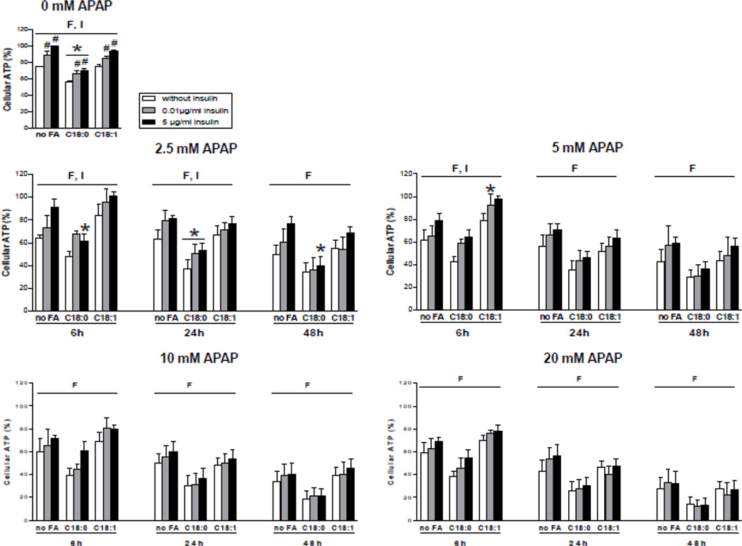
Figure 4.
Levels of ATP in HepaRG cells treated or not with APAP. HepaRG cells were incubated for 1 week with different conditions of incubation with 100 µM stearic acid (C18:0), 100 µM oleic acid (C18:1) and different concentrations of insulin and subsequently treated or not with 2.5, 5, 10 or 20 mM of APAP. ATP levels were measured 6, 24 or 48 h after APAP treatment. Cellular ATP levels were set at 100% in HepaRG cells incubated for 1 week without fatty acids and with 5 µg/ml of insulin and not treated with APAP. Results are means ± SEM for 3 to 6 independent cultures, with data in duplicates for each culture. Letters F and I indicate a significant effect (P<0.05) of fatty acids and insulin, respectively. *Significantly different from HepaRG cells incubated without fatty acids and with the same condition of insulin treatment (P<0.05). #Significantly different from HepaRG cells incubated without insulin and with the same condition of fatty acid treatment (P<0.05).

