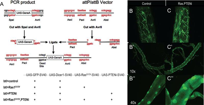
Figure 1. A multigenic Drosophila lung cancer model.
(A) Multiple UAS-containing transgenes were combined into vectors using a repeat ligation method. Up to four UAS elements were added to a single vector in the order and orientation shown. The four transgenic lines used in this study are indicated in the lower panel.
(B-C”), btl>Ras1,PTENi larvae exhibited enlarged and thickened tracheal tubes compared to btl>control larvae. Higher magnification views are shown to visualize the enlarged nuclei.

