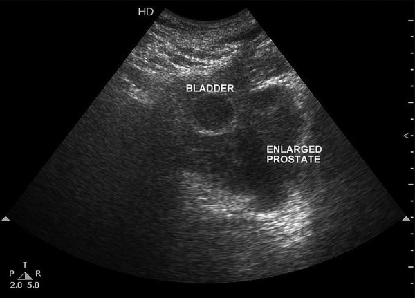
Figure 1.

Ultrasound of the pelvis in transverse view shows a heterogeneous enlarged prostate with loss of fat plane between prostate and posterior wall of the bladder, suggesting invasion of the bladder. (Bladder is empty with Foley's bulb in situ).
