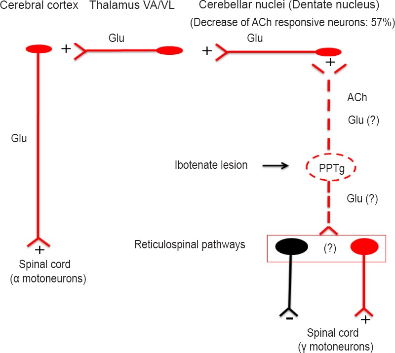
Figure 1.

Summary of the neural circuitry involved in our data.
Electrical stimulation of the PPTg activated cerebellar nuclei, likely via direct fibers. When neuronal somata in the PPTg were destroyed by ibotenic acid, a 57% decrease of activatory responses occurred in the dentate nucleus. This means that in normal condition stimulation of the PPTg would activate cerebral cortex via multiple activatory neuronal tracts. It is reasonable to hypothesize that when cholinergic neurons in the PPTg degenerate, as it occurs Parkinson's disease, the cerebral cortex becomes less active, likely involving both motor and non motor cortical areas since all the cerebellar nuclei were activated by PPTg stimulation, although at a variable extent. The loss of the PPTg input to the cerebellum would also have effects on cerebellar mechanisms involved in postural control. Finally, degeneration of PPTg neurons would also affect descending fibers to brainstem nuclei from which reticulospinal pathways arise, thus disrupting spinal cord control of muscle tone. Neuronal components: excitatory in red, inhibitory in black.
