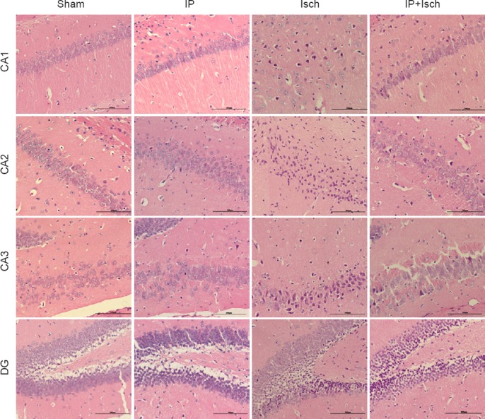
Figure 1.

IP improved the histopathology of the hippocampus following global cerebral ischemia in mice (hematoxylin-eosin staining).
Mice were subjected to histopathological assay at 24 hours post ischemia. Hematoxylin-eosin-stained hippocampal tissue from the sham, IP, Isch and IP + Isch groups (× 200). In the sham and IP groups, no obvious pathological abnormalities are observable in the hippocampal CA1–3 and DG region. Marked morphological changes are visible in the hippocampus of the Isch group, especially in the CA1–3 region; neuronal cell loss, nuclear shrinkage and dark staining of neurons are observed. Fewer damaged neurons and more normal neurons are present in these regions in the IP + Isch group, compared with the Isch group. Scale bars: 100 μm. IP: Ischemic preconditioning; Isch: ischemia; CA: cornu ammonis; DG: dentate gyrus.
