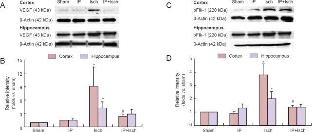
Figure 4.
IP prevented the increase in VEGF and pFlk-1 expression in the cerebral cortex and hippocampus in mice with global cerebral ischemia 24 hours after ischemia.
(A, C) Representative bands of the western blot assay for VEGF and pFlk-1 protein expression. B and D show the relative intensities of VEGF and pFlk-1. β-Actin was used as an internal control. *P < 0.05, vs. sham group; #P < 0.05, vs. Isch group. Data are expressed as the mean ± SEM from six independent animals (one-way analysis of variance followed by Bonferroni post hoc analysis). The experiment was performed in triplicate. IP: Ischemic preconditioning; Isch: ischemia; VEGF: vascular endothelial growth factor; pFlk-1: phosphorylated fetal liver kinase 1.

