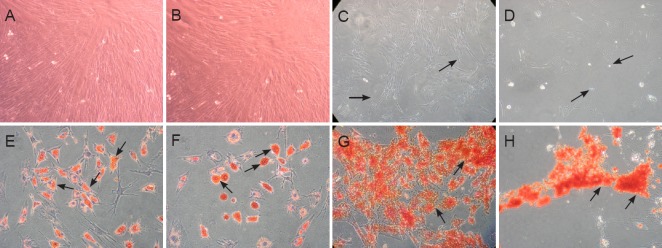
Figure 1.
Culture and identification of rat BMSCs (phase contrast microscope, × 10).
(A–D) Morphology of rat FBMSCs and MBMSCs. During subculture, attached cells exhibit a uniform fibroblast-like shape after reaching confluence at early passage. Passage 5 from female (A) and male (B) donors. Some cells from female donors (C) exhibited a heterogeneous bipolar or dendrite-like shape at late passages e.g., passage 10 (indicated by arrowheads), while cells from male donors (D) lost their normal cellular structure and gradually died (indicated by arrowheads). (E–H) Identification of BMSCs by adipogenic and osteogenic differentiation. Oil red-O staining for adipogenic differentiation shows red positive staining (indicated by arrowheads) from FBMSCs (E) and MBMSCs (F). Alizarin red staining reflects osteogenic differentiation and is shown by red positive staining (indicated by arrowheads) from FBMSCs (G) and MBMSCs (H). MBMSCs: Male bone marrow mesenchymal stem cells; FBMSCs: female bone marrow mesenchymal stem cells; BMSCs: bone marrow mesenchymal stem cells.

