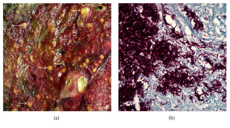
Figure 2.
(a) Macroscopic image of an area (5.5 × 5.5 cm) of the cut surface of the left lung with numerous yellowish miliary (millet seed-like) lesions, representative examples of which are arrowed. (b) Photomicrograph of a necrotic area in a hilar lymph node containing numerous acid-fast bacilli (Ziehl-Neelsen stain; magnification ×450).

